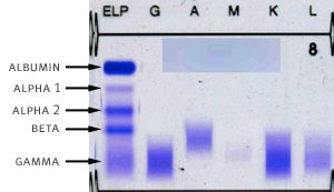
In a serum protein electrophoresis, blood serum is run through an agarose gel across an electrical current. This forces separation of the different protein components of plasma into several distinct visible bands, shown to the right.
The leftmost lane of the gel is called the serum protein electrophoresis. This lane is impregnated with a stain that will give all proteins a blue color nonspecifically. The dark band on top is albumin, one of the most common and abundant proteins in the bloodstream. The bands below each have names; alpha-1, alpha-2, beta, and gamma as marked in the figure.
Different blood components tend to aggregate in the different protein bands. For example, the HDL type of cholesterol runs in the band labeled alpha-2. All of the immunoglobulins run in the broad gamma band. The reason that the band is wide and fuzzy is that there are many different types of immunoglobulin proteins of different shapes and sizes, ranging from the small IgG subtype to the larger IgM subtype, and each runs slightly differently on the gel.
The same serum sample is run through the other lanes labeled G, A, M, K, and L shown above. These lanes constitute the immunofixation. Each of these lanes is impregnated with a specific stain that will mark only one type of protein specifically. In the "G" lane, it is the immunoglobulin type G antibody, in the "A" lane, it is the immunoglobulin type A, etc. For K and L, the stains will mark the kappa and lambda light chains respectively. In the normal human sample above, we see that the darkest bands are seen in the G and K lanes – this is because we tend to have more immunoglobulin type G and kappa light chains as compared to the other types. Still, the bands in each lane are wide and fuzzy, meaning that even within the specific subtypes of antibody, there is a variation in shape and size, because different molecules are produced by a variety of different plasma cells normally.

In the serum protein electrophoresis/immunofixation from a person with a monoclonal paraprotein (shown to the right), the gamma band is narrowed and sharp. There is now one predominant kind of immunoglobulin (called the monoclonal immunoglobulin) that runs at one precise spot on the gel, giving rise to the famous M-spike. By looking at the immunofixation lanes, we can easily see a prominent sharp band in the M and L lanes. This signifies that in this care, the M-protein produced is the IgM-lambda subtype. This M-spike can be measured and followed throughout the clinical course of an individual and roughly reflects the amount of malignant plasma cells in the body. A rapidly rising M-protein connotes aggressive growing myeloma, while falling or disappearing M-protein can signify a good response to treatment.